On the anterior and posterior views of the muscular system above, superficial muscles (those at the surface) are shown on the right side of the body while deep muscles (those underneath the superficial muscles) are shown on the left half of the body.
Muscle Diagram Most Important Muscles Of An Athletic Male Body Anterior And Posterior View Labeled Isolated Vector Illustration On White Background Stock Illustration – Download Image Now – iStock
externus. outside. EXternal. internus. inside. INternal. Anatomists name the skeletal muscles according to a number of criteria, each of which describes the muscle in some way. These include naming the muscle after its shape, its size compared to other muscles in the area, its location in the body or the location of its attachments to the

Source Image: coursehero.com
Download Image
We are given a picture of a muscle tissue and we are to label the parts of it. Let’s start with the first one. Alright. The first box depends on something. … Correctly label the following muscles of the posterior view: – Flexor hallucis longus – Lateral rotators – Tibialis posterior – Iliotibial band – Serratus anterior – Supraspinatus

Source Image: ahajournals.org
Download Image
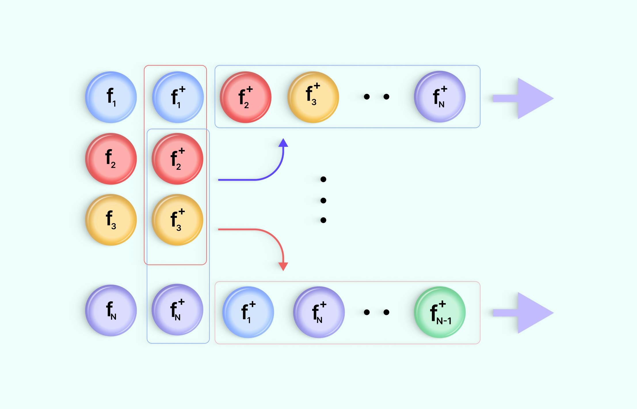
Full Guide to Contrastive Learning | Encord Jan 17, 2023The superficial muscles of the back are responsible for movement of the shoulder. The intermediate muscles of the back assist in the movement of the rib cage during respiration. The intrinsic back muscles facilitate movement of the head and neck and are fundamental in maintaining posture and balance. The posterior or back muscles perform a wide

Source Image: centerforvein.com
Download Image
Correctly Label The Following Muscles Of The Posterior View
Jan 17, 2023The superficial muscles of the back are responsible for movement of the shoulder. The intermediate muscles of the back assist in the movement of the rib cage during respiration. The intrinsic back muscles facilitate movement of the head and neck and are fundamental in maintaining posture and balance. The posterior or back muscles perform a wide VIDEO ANSWER: We have to show the structures of the shoulder and parlymso in the given figure. The head of numerous, the head of humorous, and the head of the sergical neck are the first living things. Further, further. The scapula is the third one.
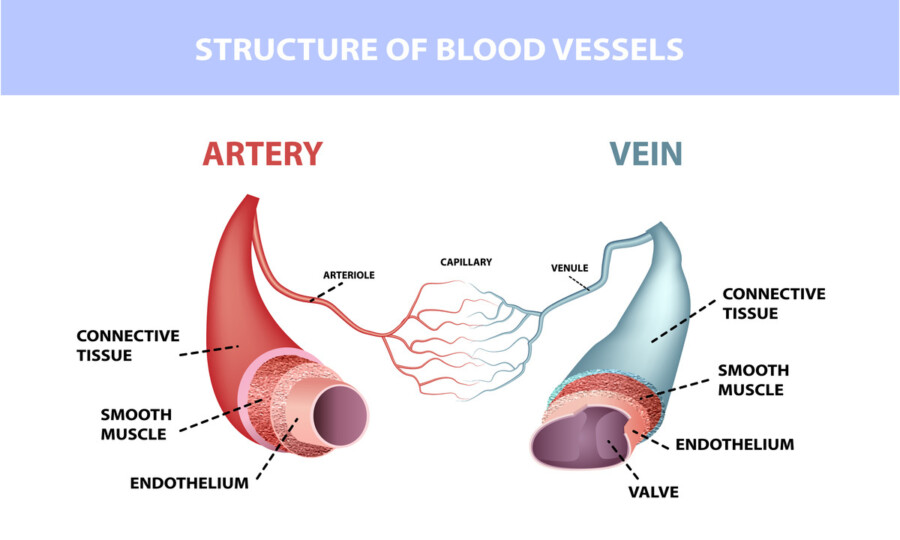
Arteries and Veins: What’s the Difference?
Correctly label the following muscles of the posterior view. Semitendinosus Serratus posterior inferior Semispinalis capitis Extensor carpi ulnaris Triceps brachii Trapezius Teres minor External abdominal oblique This problem has been solved! You’ll get a detailed solution from a subject matter expert that helps you learn core concepts. See Answer 7 Health Benefits Of Lunges – PharmEasy Blog

Source Image: pharmeasy.in
Download Image
Emotion AI: How can AI understand Emotions? – Twine Blog Correctly label the following muscles of the posterior view. Semitendinosus Serratus posterior inferior Semispinalis capitis Extensor carpi ulnaris Triceps brachii Trapezius Teres minor External abdominal oblique This problem has been solved! You’ll get a detailed solution from a subject matter expert that helps you learn core concepts. See Answer

Source Image: twine.net
Download Image
Muscle Diagram Most Important Muscles Of An Athletic Male Body Anterior And Posterior View Labeled Isolated Vector Illustration On White Background Stock Illustration – Download Image Now – iStock On the anterior and posterior views of the muscular system above, superficial muscles (those at the surface) are shown on the right side of the body while deep muscles (those underneath the superficial muscles) are shown on the left half of the body.

Source Image: istockphoto.com
Download Image
Full Guide to Contrastive Learning | Encord We are given a picture of a muscle tissue and we are to label the parts of it. Let’s start with the first one. Alright. The first box depends on something. … Correctly label the following muscles of the posterior view: – Flexor hallucis longus – Lateral rotators – Tibialis posterior – Iliotibial band – Serratus anterior – Supraspinatus

Source Image: encord.com
Download Image
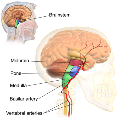
Brainstem – Physiopedia Muscle enabling the hand to extend and to draw near the median axis of the body. Muscle enabling all the fingers, except the thumb, to extend; it also helps the hand to extend on the forearm. Short muscle reinforcing the action of the triceps; it allows the forearm to extend on the arm and also stabilizes the elbow joint. Muscle enabling the

Source Image: physio-pedia.com
Download Image
Megakaryocytes and platelet-fibrin thrombi characterize multi-organ thrombosis at autopsy in COVID-19: A case series – eClinicalMedicine Jan 17, 2023The superficial muscles of the back are responsible for movement of the shoulder. The intermediate muscles of the back assist in the movement of the rib cage during respiration. The intrinsic back muscles facilitate movement of the head and neck and are fundamental in maintaining posture and balance. The posterior or back muscles perform a wide

Source Image: thelancet.com
Download Image
17E60840-5AD9-448F-B7D2-E1DB70FD4769.jpeg – Correctly label the following muscles of the anterior view . Superficial | Deep Tibialis | Course Hero VIDEO ANSWER: We have to show the structures of the shoulder and parlymso in the given figure. The head of numerous, the head of humorous, and the head of the sergical neck are the first living things. Further, further. The scapula is the third one.

Source Image: coursehero.com
Download Image
Emotion AI: How can AI understand Emotions? – Twine Blog
17E60840-5AD9-448F-B7D2-E1DB70FD4769.jpeg – Correctly label the following muscles of the anterior view . Superficial | Deep Tibialis | Course Hero externus. outside. EXternal. internus. inside. INternal. Anatomists name the skeletal muscles according to a number of criteria, each of which describes the muscle in some way. These include naming the muscle after its shape, its size compared to other muscles in the area, its location in the body or the location of its attachments to the
Full Guide to Contrastive Learning | Encord Megakaryocytes and platelet-fibrin thrombi characterize multi-organ thrombosis at autopsy in COVID-19: A case series – eClinicalMedicine Muscle enabling the hand to extend and to draw near the median axis of the body. Muscle enabling all the fingers, except the thumb, to extend; it also helps the hand to extend on the forearm. Short muscle reinforcing the action of the triceps; it allows the forearm to extend on the arm and also stabilizes the elbow joint. Muscle enabling the